A biomedical imaging technique capable of capturing mechanical properties of bodily tissue could improve diagnosis and treatment of breast cancer and other diseases.
XSEDE is helping advance the imaging technology, called NLACE, with high-performance computing and a Web-based science gateway that lets researchers submit data easily and get results automatically. The research is a multi-institutional effort led by Paul Barbone at Boston University and Assad Oberai at Rensselaer Polytechnic Institute.
Numerous pathologies are linked to changing mechanical properties of tissue, from swollen glands and hardening of the arteries to cirrhosis of the liver and stiff lumps that can signal breast or other cancers.Removing and testing tissue samples can determine mechanical property changes, but NLACE promises precise measurement without invasive sample taking, says Barbone, a professor of theoretical acoustics and applied mechanics.
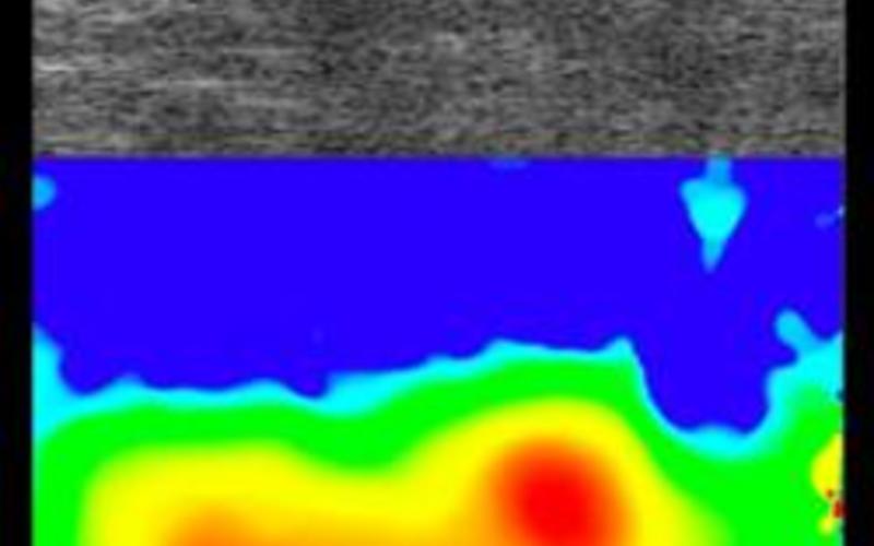
Barbone likens coupling NLACE with ultrasound, magnetic resonance imaging (MRI) and other commonly used biomedical imaging technologies to adding infrared capability to a regular camera. Through the additional information NLACE generates about tissue mechanical properties, it can show things, like a budding tumor, that might be invisible to standard scanning methods alone.
Clinical methods called elastography already can noninvasively measure linear elastic properties at millimeter scale in soft tissues, capturing how tissue reacts to gentle manipulation by ultrasound, MRI and other imaging methods. But NLACE’s power is a flexibility that allows it to capture much more.
“NLACE has been applied to soft tissues like organs and hard tissues like bone; it’s even measured mechanical property distributions within individual living cells,” Barbone says. “NLACE can infer not just linear, but also nonlinear properties; not just properties that are the same in any direction, but also directionally dependent properties that vary depending on the direction the material is pulled; not just how elastic the tissue is, but how fluid and viscous as well.”
Besides potential for integrating NLACE into clinical scanners, the research advances basic imaging science, along with understanding of how tissue mechanical properties play into development of diseases. In breast cancer, for instance, evidence exists that mechanical property changes may actually spur malignancy, Barbone notes.
“We used to think only that some diseases cause a change in the mechanical properties,” Barbone says. “Now we recognize that in some cases, changes in mechanical properties contribute to the disease. The mechanical properties of tissues surrounding a tumor, for example, can influence the biological behavior of tumor cells.”
The research to develop NLACE requires high-performance computing. Essentially, the researchers are presented with a result and then have to computationally reverse engineer an optimal answer to why they got it, says Oberai, a professor and associate director of Rensselaer Polytechnic’s Scientific Computation Research Center.
They run problems through hundreds, even thousands, of iterations to glean optimal solutions. Without XSEDE supercomputers like Gordon at the San Diego Supercomputer Center (SDSC) at the University of California, San Diego, the time to do so would be too great. Gordon, which is designed for data-intensive work, also has large memory capacity, which makes it ideal for “a big data problem” like the NLACE research, Oberai says.